
The Ultrasound Laboratory is an academic research group dedicated to advancing medical ultrasound, with a core mission of translational research and the development of novel ultrasound imaging and therapeutic technologies. By bridging fundamental acoustic science with rigorous clinical application, our multidisciplinary team aims to enhance diagnostic precision, improve image-guided therapies, and ultimately elevate patient care.
Research focus areas
Microbubble image-guided vascular therapy
We are developing ultrasound-based, image-guided vascular therapies. By combining microbubble contrast agents with high-frequency microvascular imaging and therapeutic ultrasound, we aim to optimize interventions, such as ischemic therapy and hyperthermia. This integrated approach allows for simultaneous imaging and targeted treatment.
Quantitative and computational sonography
A central goal of the laboratory is to improve the precision and reproducibility of ultrasound through automation and quantification. We leverage advanced computational techniques, including segmentation, feature analysis, and machine learning, applied to B-mode and Doppler imaging. Our work in ultrasound radiomics, echo statistics, and texture analysis is actively being applied to the early detection and assessment of hepatic fibrosis.
Photoacoustic molecular imaging
The lab is at the forefront of developing photoacoustic imaging (PAI) for endogenous molecular targets, including melanin, oxygenation, and collagen. Our recent efforts include utilizing spectral PAI and superpixel unmixing analysis for collagen tracking, as well as assessing tissue oxygenation in models of hepatic fibrosis, offering new avenues for high-resolution, non-invasive molecular characterization.
Oncology and image-guided interventions
We are actively engaged in improving the early detection and targeted treatment of cancer, specifically hepatocellular carcinoma (HCC). Our research includes using hydralazine to enhance ultrasound image contrast for early HCC detection and to develop image-guided therapeutic strategies based on vasomodulation.
Cardiovascular research: ultrasound endothelial diagnostic platform
Our laboratory is actively developing a non-invasive ultrasound-based endothelial diagnostic platform to detect and characterize vascular dysfunction at its earliest, subclinical stages. By integrating high-resolution B-mode and Doppler imaging with our established expertise in vascular imaging and machine learning, we aim to provide sensitive, reproducible metrics of endothelial health. Ultimately, this research seeks to translate comprehensive endothelial assessment into a practical point-of-care diagnostic instrument for earlier cardiovascular risk stratification.
Collaborative and translational philosophy
Translation is central to the Ultrasound Lab's mission. Our progress is built on extensive collaboration with a diverse network of clinical and scientific experts across radiology, veterinary medicine, physics, and bioengineering. We work closely with our collaborators to ensure that our innovations are rigorously evaluated in preclinical models and optimized for clinical workflows, transforming benchtop discoveries into tangible medical technologies.
Laboratory details
Leadership
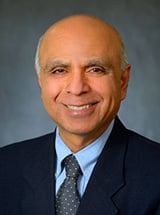 Sandy Sehgal, PhD
Professor, Director of Ultrasound Research
Staff
- Mrigendra Karmacharya, PhD
Senior Scientist - Theodore Cary, MS
Computer Scientist - Laith Sultan, MD MPH
Collaborator - Hersh Sagreiya, MD
Collaborator (Radiology) - Angela Brice, DVM PhD
Collaborator (Pathology) - Quy Xuan Cao, PhD
Collaborator (Biostatistics) - Rachel Alisio
Administrative Assistant
Liver Ultrasound
- Sultan LR, Venkatakrishna SSB, Anupindi SA, Andronikou S, Acord MR, Otero HJ, Darge K, Sehgal CM, Holmes JH. ChatGPT-4-Driven Liver Ultrasound Radiomics Analysis: Diagnostic Value and Drawbacks in a Comparative Study. JMIR AI 4: e68144, Jun 2025. PMCID: PMC12260471. PMID: 40388838
- Sultan LR, Karmacharya MB, Al-Hasani M, Cary TW, Sehgal CM. Hydralazine-augmented contrast ultrasound imaging improves the detection of hepatocellular carcinoma. Med Phys 2023. PMCID: PMC10128060
- Al-Hasani M, Sultan LR, Sagreiya H, Cary TW, Karmacharya MB, Sehgal CM. Ultrasound Radiomics for the Detection of Early-Stage Liver Fibrosis. Diagnostics (Basel). 2022 Nov 9;12(11):2737. doi: 10.3390/diagnostics12112737. PMID: 36359580
- Sultan LR, Cary TW, Al-Hasani M, Karmacharya MB, Venkatesh SS, Assenmacher CA, Radaelli E and Sehgal CM. Can Sequential Images from the Same Object Be Used for Training Machine Learning Models? A Case Study forDetecting Liver Disease by Ultrasound Radiomics. AI 3: 739-750, Sep 2022. PMID: 36168560, PMCID: PMC9511699, DOI: 3390/ai3030043
- Machado P, Gupta I, Gummadi S, Stanczak M, Wessner CE, Fenkel JM, Shaw CM, Shamini-Noori S, Schultz S, Soulen MC, Sehgal CM, Wallace K, Eisenbrey JR, Forsberg F.: Hepatic Vein Contrast-Enhanced Ultrasound Subharmonic Imaging Signal as a Screening Test for Portal Hypertension. Dig Dis Sci 66(12): 4354-4360, Dec 2021 Notes: DOI: 10.1007/s10620-020-06790-6.
- Chauhan A, Shamchi SP, Dumon KR, Williams NN, Sehgal CM: Reliability of acoustic radiation force impulse shear wave elastography in the evaluation of liver stiffness in morbidly obese patients. J Clin Ultrasound 48(7): 369-376, Sep 2020. PMID: 32491197, DOI: 1002/jcu.22837
- D'Souza Julia C, Sultan Laith R, Hunt Stephen J, Schultz Susan M, Brice Angela K, Wood Andrew K W, Sehgal Chandra M: B-mode ultrasound for the assessment of hepatic fibrosis: a quantitative multiparametric analysis for a radiomics approach. Scientific Reports 9(1): 8708, Jun 2019 Notes: DOI: 1038/s41598-019-45043-z. PMCID: PMC6581954
- Chauhan A, Sultan LR, Furth EE, Jones LP, Khungar V, Sehgal CM.: Diagnostic Accuracy of Hepatorenal Index in the Detection and Grading of Hepatic Steatosis. Journal of Clinical Ultrasound 44(9): 580-586, Nov 2016 Notes: DOI: 1002/jcu.22382.
- Sehgal C M: Quantitative relationship between tissue composition and scattering of ultrasound. The Journal of the Acoustical Society of America 94(4): 1944-1952, Oct 1993 Notes: DOI: 1121/1.407517.
- Sehgal C M, Brown G M, Bahn R C, Greenleaf J F: Measurement and use of acoustic nonlinearity and sound speed to estimate composition of excised livers. Ultrasound in Medicine & Biology 12(11): 865-874, Nov 1986 Notes: DOI: 1016/0301-5629(86)90004-9.
Breast Ultrasound
- Moustafa AF, Cary TW, Sultan LR, Schultz SM, Conant EF, Venkatesh SS, Sehgal CM: Color Doppler Ultrasound Improves Machine Learning Diagnosis of Breast Cancer. Diagnostics 107(9), Aug 2020.
- Wu T, Sultan LR, Tian J, Cary TW, Sehgal CM: Machine learning for diagnostic ultrasound of triple-negative breast cancer. Breast Cancer Research and Treatment 173(2): 365-373, Jan 2019 Notes: DOI: 10.1007/s10549-018-4984-7. PMCID: 30343454
- Venkatesh SS, Levenback BJ, Sultan LR, Bouzghar G, Sehgal CM: Going beyond a first reader: A machine learning methodology for optimizing cost and performance in breast ultrasound diagnosis. Ultrasound in Medicine & Biology 41(12): 3148-3162, Dec 2015 Notes: DOI: 1016/j.ultrasmedbio.2015.07.020.
- Sultan LR, Bouzghar G, Levenback BJ, Nauroze Faizi A, Santosh S. Venkatesh SS, Conant EF, Sehgal CM: Observer Variability in BI-RADS Ultrasound Features and Its Influence on Computer-Aided Diagnosis of Breast Masses. Advances in Breast Cancer Research 4(1): 1-8, Jan 2015 Notes: DOI: 4236/abcr.2015.41001.
- Bouzghar G, Levenback BJ, Sultan LR, Venkatesh SS, Cwanger A, Conant EF, Sehgal CM: Bayesian probability of malignancy with BI-RADS sonographic features. Journal of Ultrasound in Medicine 33(4): 641-648, Apr 2014 Notes: DOI: 7863/ultra.33.4.641.
- Weinstein Susan P, Conant Emily F, Sehgal Chandra M, Woo Irene P, Patton Jill A: Hormonal variations in the vascularity of breast tissue. Journal of Ultrasound in Medicine 24(1): 67-72, Jan 2005 Notes: DOI: 7863/jum.2005.24.1.67.
- Weinstein SP, Sehgal CM, Conant EF, Patton JA: Micro-calcification in breast tissue phantoms visualized with acoustic resonance coupled with power Doppler US: Initial observations. Radiology 224(1): 265-269, Jul 2002 Notes: DOI: 1148/radiol.2241010511.
- Arger P H, Sehgal C M, Conant E F, Zuckerman J, Rowling S E, Patton J A: Interreader variability and predictive value of US descriptions of solid breast masses: pilot study. Academic Radiology 8(4): 335-342, Apr 2001 Notes: DOI: 10.1016/S1076-6332(03)80503-2.
Lung Ultrasound
- Sultan LR, Haertter A, Al-Hasani M, Demiris G, Cary TW, Tung-Chen Y, Sehgal CM.: Can Artificial Intelligence Aid Diagnosis by Teleguided Point-of-Care Ultrasound? A Pilot Study for Evaluating a Novel Computer Algorithm for COVID-19 Diagnosis Using Lung Ultrasound. AI (Basel) 2023. PMCID: PMC10623579
- Sultan LR, Chen YT, Cary TW, Ashi K, Sehgal CM. Quantitative pleural line characterization outperforms traditional lung texture ultrasound features in detection of Covid-19. JACEP 2021.
- Sultan LR, Chen YT, Cary TW, Ashi K, Sehgal CM.: Quantitative pleural line characterization outperforms traditional lung texture ultrasound features in detection of COVID-19. J Am Coll Emerg Physicians Open 2: e12418, Apr 2021. PMCID: PMC8018308
Musculoskeletal Ultrasound
- Newton JB, Nuss CA, Weiss SN, Betts RL, Sehgal CM, Soslowsky LJ.: In Vivo Photoacoustic Ultrasound (PAUS) Assay for Monitoring Tendon Collagen Compositional Changes during Injury and Healing. Diagnostics (Basel) 14: 1498, Jul 2024. PMCID: PMC11275422
- Riggin CN, Weiss SN, Rodriguez AB, Raja H, Chen M, Schultz SM, Sehgal CM, Soslowsky LJ.: Increasing Vascular Response to Injury Improves Tendon Early Healing Outcome in Aged Rats. Ann Biomed Eng 2022. PMCID: PMC9107615
- Riggin CN, Rodriguez AB, Weiss SN, Raja HA, Chen M, Schultz SM, Sehgal CM, Soslowsky LJ.: Modulation of vascular response after injury in the rat Achilles tendon alters healing capacity. J Orthop Res 39(9): 2000-2016, Sep 2021 Notes: DOI: 10.1002/jor.24861. PMCID: PMC7960560
- Riggin Corinne N, Schultz Susan M, Sehgal Chandra M, Soslowsky Louis J: Ultrasound Evaluation of Anti-Vascular Endothelial Growth Factor-Induced Changes in Vascular Response Following Tendon Injury. Ultrasound in Medicine & Biology 45(7): 1841-1849, Jul 2019 Notes: DOI: 1016/j.ultrasmedbio.2019.03.00. PMCID: PMC6555687
Tongue Ultrasound
- Parekh MH, Thuler E, Triantafillou V, Seay E, Sehgal C, Schultz S, Keenan BT, Schwartz AR, Dedhia RC.: Physiologic and anatomic determinants of hyoid motion during drug-induced sleep endoscopy. Sleep Breath Jul 2024.
- Parekh MH, Thuler E, Triantafillou V, Seay E, Sehgal C, Schultz S, Keenan BT, Schwartz AR, Dedhia RC.: The Application of Ultrasound to Quantify Hyoid Motion During Drug-Induced Sleep Endoscopy. Laryngoscope 2023. PMCID: PMC10592545
- Yu JL, Wiemken A, Schultz SM, Keenan BT, Sehgal CM, Schwab RJ.: A comparison of ultrasound echo intensity to magnetic resonance imaging as a metric for tongue fat evaluation. Sleep 45: zsab295, Feb 2022. PMCID: PMC8842321
Cardiovascular Ultrasound
- Sidnawi B, Zhou B, Chen Z, Sehgal C, Santhanam S, Wu Q.: A comprehensive physics-based model for the brachial Artery's full flow mediated dilation (FMD) response observed during the FMD test. Comput Biol Med 2024. PMCID: PMC11324374
- Sidnawi B, Chen Z, Sehgal C, Santhanam S, Wu Q.: On the modeling of mechanotransduction in flow-mediated dilation. J Mech Behav Biomed Mater 120: 104606, Aug 2021 Notes: DOI: 10.1016/j.jmbbm.2021.104606.
- Sidnawi B, Chen Z, Sehgal C, Wu Q: Characterization of arterial flow mediateddilation via a physics-based model. J Mech Behav Biomed Mater. 107: 103756, Jul 2020.
- Chen Z, Sultan LR , Schultz SM,Cary TW and Sehgal CM: Brachial flow-mediated dilation by continuous monitoring of arterial cross-section with ultrasound imaging. Ultrasound 27(4): 241-251, Nov 2019 Notes: DOI: 1177/1742271X19857770.
- Pugliese DN, Sehgal CM, Sultan LR, Reamer CB, Mohler ER 3rd. : Feed-forward active contour analysis for improved brachial artery reactivity testing. Vascular Medicine 21(4): 317-324, Aug 2016 Notes: DOI: 1177/1358863X16634194.
- Pouch AM. Yushkevich PA. Jackson BM. Jassar AS. Vergnat M. Gorman JH. Gorman RC. Sehgal CM : Development of a semi-automated method for mitral valve modeling with medial axis representation using 3D ultrasound. Medical Physics 39(2): 933-950, Feb 2012 Notes: DOI: 1118/1.3673773. PMCID: PMC3281970
- Eraso LH, Reilly MP, Sehgal C,Mohler ER 3rd. : Emerging diagnostic and therapeutic molecular imaging applications in vascular disease. Vascular Medicine 16(2): 145-156, Apr 2011 Notes: DOI: 1177/1358863X10392474. PMCID: PMC3750985
- Mohler Emile R, Sibley Alexandra A, Schultz Susan M, Zhang Lifeng, Sehgal Chandra M: High-frequency ultrasound for evaluation of intimal thickness. Journal of the American Society of Echocardiography 22(10): 1129-1133, Oct 2009 Notes: DOI: 1016/j.echo.2009.06.021. PMCID: PMC2762201
- Mohler Emile R, Sehgal Chandra M, Ferrari Victor A, Parmacek Michael, Shih Anthony, Wilensky Robert L: A novel ultrasound method for evaluation of collateral development in limb ischemia. Vascular Medicine 7(3): 169-175, Aug 2002 Notes: DOI: 1191/1358863x02vm431oa.
- Mohler E R, Klugherz B, Goldman R, Kimmel S E, Wade M, Sehgal C M: Trial of a novel prostacyclin analog, UT-15, in patients with severe intermittent claudication. Vascular Medicine 5(4): 231-237, Nov 2000.
- Shlansky-Goldberg R D, Cines D B, Sehgal C M: Catheter-delivered ultrasound potentiates in vitro thrombolysis. Journal of Vascular and Interventional Radiology 7(3): 313-320, May-Jun 1996 Notes: DOI: 1016/s1051-0443(96)72861-0.
- Kao Yen Hong, Mohler Emile R, Arger Peter H, Sehgal Chandra M: Brachial artery: measurement of flow-mediated dilatation with cross-sectional US--technical validation. Radiology 228(3): 895-900, Sep 2003 Notes: DOI: 1148/radiol.2283020966.
Photoacoustic
- Sultan LR, Grasso V, Jose J, Al-Hasani M, Karmacharya MB, Sehgal CM.: Advanced Techniques for Liver Fibrosis Detection: Spectral Photoacoustic Imaging and Superpixel Photoacoustic Unmixing Analysis for Collagen Tracking. Sensors (Basel) 24: 4617, Jul 2024. PMCID: PMC11281248.
- Kirkham Brooke M, Schultz Susan M, Ashi K, Sehgal Chandra M: Assessment of Age-related Oxygenation Changes in Calf Skeletal Muscle by Photoacoustic Imaging: A Potential Tool for Peripheral Arterial Disease. Ultrasonic Imaging 41(5): 290-300, Sept 2019 Notes: DOI: 1177/0161734619862287.
- Karmacharya MB, Sultan LR, Sehgal CM. Photoacoustic monitoring of oxygenation changes induced by therapeutic ultrasound in murine hepatocellular carcinoma. Sci Rep. 2021 Feb 18;11(1):4100. doi: 10.1038/s41598-021-83439-y. PubMed PMID: 33603035; PubMed Central PMCID: PMC7893035.
- Karmacharya MB, Sultan LR, Kirkham BM, Brice AK, Wood AKW, Sehgal CM.: Photoacoustic Imaging for Assessing Tissue Oxygenation Changes in Rat Hepatic Fibrosis. Diagnostics (Basel) 10: 705, Sep 2020. PMCID: PMC7555416
- Chen Z, Chattaraj R, Pulsipher KW, Karmacharya MB, Hammer DA, Lee D, Sehgal CM.: Photoacoustic and Ultrasound Dual-Mode Imaging via Functionalization of Recombinant Protein-Stabilized Microbubbles with Methylene Blue. ACS Appl Bio Mater 2: 4020-4026, Sep 2019.
- Sinnamon Andrew J, Neuwirth Madalyn G, Song Yun, Schultz Susan M, Liu Shujing, Xu Xiaowei, Sehgal Chandra M, Karakousis Giorgos C: Multispectral photoacoustic imaging for the detection of subclinical melanoma. Journal of Surgical Oncology 119(8): 1070-1076, Jun 2019 Notes: DOI: 1002/jso.25447
Therapeutic antivascular ultrasound
- Moradpour M, Li Z, Al-Daoud O, Yazdanpanah F, Cheruvu A, Sehgal C, Hunt S.: Efficacy of Antivascular Ultrasound (AVUS) in Hepatocellular Carcinoma (HCC). Cancers (Basel) 16: 3756, Nov 2024. PMCID: PMC11592062
- Morrison RG, Karmacharya MB, Sehgal CM.: Contrast-Enhanced Ultrasound Tumor Therapy With Abdominal Imaging Transducer. J Med Device 16: 041010, Dec 2022. PMCID: PMC9445316
- Karmacharya MB, Sultan LR, Hunt SJ, Sehgal CM.: Hydralazine augmented ultrasound hyperthermia for the treatment of hepatocellular carcinoma. Sci Rep 11: 15553, Jul 2021. PMCID: PMC8324788
- Sultan LR, Karmacharya MB, Hunt SJ, Wood AKW, Sehgal CM. Subsequent Ultrasound Vascular Targeting Therapy of Hepatocellular Carcinoma Improves the Treatment Efficacy. Biology (Basel). 2021 Jan. 22;10(2)
- D'Souza JC, Sultan LR, Hunt SJ, Gade TP, Karmacharya MB, Schultz SM, Brice AK, Wood AKW, Sehgal CM. Microbubble-enhanced ultrasound for the antivascular treatment and monitoring of hepatocellular carcinoma. Nanotheranostics. 2019;3(4):331-341.
- DeOre Brandon J, Galie Peter A, Sehgal Chandra M: Fluid flow rate dictates the efficacy of low-intensity antivascular ultrasound therapy in a microfluidic model. Microcirculation 26(7): e12576, Oct 2019 Notes: DOI: 1111/micc.12576. PMCID: PMCID: PMC6819214
- Hunt SJ, Gade T, Pickup S, Soulen MC, Seghal C. Antivascular ultrasound therapy: magnetic resonance imaging validation and activation of the immune response in murine melanoma. J Ultrasound Med. 2015 Feb;34(2):275-87.
- Wood Andrew K W, Bunte Ralph M, Schultz Susan M, Sehgal Chandra M: Acute increases in murine tumor echogenicity after antivascular ultrasound therapy: a pilot preclinical study. Journal of Ultrasound in Medicine 28(6): 795-800, Jun 2009 Notes: DOI: 7863/jum.2009.28.6.795.Wood
- Andrew K W, Bunte Ralph M, Price Heather E, Deitz Margaret S, Tsai Jeff H, Lee William M-F, Sehgal Chandra M: The disruption of murine tumor neovasculature by low-intensity ultrasound-comparison between 1- and 3-MHz sonication frequencies. Academic Radiology 15(9): 1133-1141, Sep 2008 Notes: DOI: 1016/j.acra.2008.04.012. PMCID: PMC2597509Wood Andrew K W,
Ultrasound techniques and algorithms
- Ashi KH, Karmacharya MB, Sultan LR, Guerrero DT, Chorny M,Sehgal CM: Photoacoustic imaging of vascular hyperpermeability. IEEE International Ultrasonics Symposium 2023. IEEE Xplore, October 2023 Notes: DOI: 10.1109/IUS51837.2023.10307871.
- Ashi K, Kirkham B, Chauhan A, Schultz SM, Brake BJ, Sehgal CM.: Quantitative colour Doppler and greyscale ultrasound for evaluating prostate cancer. Ultrasound 29(2): 106-111, May 2021 Notes: DOI: 10.1177/1742271X20952825. PMCID: PMC8083141
- Chattaraj R, Hwang M, Zemerov SD, Dmochowski IJ, Hammer DA, Lee D, Sehgal CM: Ultrasound Responsive Noble Gas Microbubbles for Applications in Image-Guided Gas Delivery. Adv Healthc Mater 9(9): e1901721, May 2020. PMCID: PMC7457952
- Chen Zhuo, Pulsipher Katherine W, Chattaraj Rajarshi, Hammer Daniel A, Sehgal Chandra M, Lee Daeyeon: Engineering the Echogenic Properties of Microfluidic Microbubbles Using Mixtures of Recombinant Protein and Amphiphilic Copolymers. Langmuir : the ACS journal of surfaces and colloids 35(31): 10079-10086, Aug 2019 Notes: DOI: 1021/acs.langmuir.8b03882. PMCID: PMC6698903
- Schwartz N, Siegal J, Rourke A, Sehgal CM. Placental Pulsatility: Quantitative Assessment of Placental Bed Vasculature by 2-Dimensional Doppler Cine Imaging. J Ultrasound Med. 2019 Feb;38(2):471-479.
- Pulsipher KW, Hammer DA, Lee D, Sehgal CM.: Engineering Theranostic Microbubbles Using Microfluidics for Ultrasound Imaging and Therapy: A Review. Ultrasound Med Biol 44(12): 2441-2460, Dec. 2018. PMCID: 6643280
- Sultan LR, Xiong H, Zafar HM, Schultz SM, Langer JE, Sehgal CM. Vascularity assessment of thyroid nodules by quantitative color Doppler ultrasound. Ultrasound Med Biol. 2015 May;41(5):1287-93. 2015
- Cary TW, Reamer CB, Sultan LR, Mohler ER 3rd, Sehgal CM: Brachial artery vasomotion and transducer pressure effect on measurements by active contour segmentation on ultrasound. Medical Physics 41(2): 022901, Feb 2014 Notes: DOI: 1118/1.4862508. PMCID: PMC3987701
- Pouch Alison M, Cary Theodore W, Schultz Susan M, Sehgal Chandra M: In vivo noninvasive temperature measurement by B-mode ultrasound imaging. Journal of Ultrasound in Medicine 29(11): 1595-1606, Nov 2010 Notes: DOI: 7863/jum.2010.29.11.1595.
- Sehgal Chandra M, Cary Theodore W, Arger Peter H, Wood Andrew K W: Delta-projection imaging on contrast-enhanced ultrasound to quantify tumor microvasculature and perfusion. Academic Radiology 16(1): 71-78, Jan 2009 Notes: DOI: 1016/j.acra.2008.07.013. PMCID: PMC2644424
- Cary T W, Conant E F, Arger P H, Sehgal C M: Diffuse boundary extraction of breast masses on ultrasound by leak plugging. Medical physics 32(11): 3318-3328, Nov 2005 Notes: DOI: 1118/1.2012967
- Kamotani Yoko, Lee William M F, Arger Peter H, Cary Theodore W, Sehgal Chandra M: Multigated contrast-enhanced power Doppler to measure blood flow in mice tumors. Ultrasound in Medicine & Biology 29(7): 977-984, Jul 2003 Notes: DOI: 1016/s0301-5629(03)00054-1.
Patents
Calcification:
- Sehgal CM. Apparatus for imaging an element within a mass and method therefore for detection of breast macrocalcifications. 5,997,477
3D imaging:
- Gorman JH, Pouch AM, Gorman RC, Wang H, Yushkevich P, Jackson BM, Avants BB, Sehgal CM. Fully automated image segmentation of heart valves using multi-atlas label fusion and deformable medial modeling. US 9,406,142.
- Jackson BM, Gorman RC, Gorman JH, Pouch AM, Sehgal CM, Yushkevich P, Avants BB Wang H, B,. Semi-automated heart valve morphometry and computational stress analysis from 3D images US 10,783,631.
Microbubbles:
- Sehgal CM: Method and Apparatus to Control to Control Microbubble destruction during Contrast-enhanced Ultrasound Imaging. And Uses Therefore. US 6,858,011;
- Lee D, Sehgal CM, Lee Microfluidic-based generation of functional microbubbles for ultrasound imaging and therapy. US 20120328529;
- Lee D, Angile F, Vargo K, Hammer DA, Sehgal CM. Apparatus and methods for making recombinant protein-stabilized monodisperse microbubbles. US20170119911
Diagnostic AI:
- Sehgal CM, Venkatesh SS, Sultan LR. Machine implemented methods, systems, and apparatuses for improving diagnostic performance. US2017014124.
Chandra (Sandy) M. Sehgal, PhD
Professor, Director of Ultrasound Research
Department of Radiology
3620 Hamilton Walk, Office 168B
Philadelphia, PA 19104
Office Phone: 215-349-5461
Lab Phone: 215-662-6673
Fax: 215-898-6115
Email: Chandra.Sehgal@pennmedicine.upenn.edu
Jill E. Langer, MD
US Section Chief
Program Director, Abdominal Imaging Fellowship
Professor of Radiology
Department of Radiology
3400 Spruce Street
Philadelphia, PA 19104
Phone: 215-662-7012
Email: Jill.Langer@pennmedicine.upenn.edu
Susan M. Schultz, RDMS
Department of Radiology
3620 Hamilton Walk, Office 168A
Philadelphia, PA 19104
Phone: 215-573-0972
Fax: 215-898-6115
Email: Susan.Schultz@pennmedicine.upenn.edu