
Focus
Our mission is to develop and translate noninvasive MR- and PET-based metabolic imaging tools that reveal actionable metabolic vulnerabilities and accelerate the development of new therapies. We integrate rigorous molecular and cellular validation with preclinical animal studies and matched co-clinical trials, advancing promising imaging biomarkers through IND-enabling steps and into patient studies through close partnerships with physician-scientists and industry.
Leadership
 Rong Zhou, PhD
Director of TMMI Lab
Contact us
198 John Morgan Building
3620 Hamilton Walk
Phone: (215) 746-8747
Email: rongzhou@pennmedicine.upenn.edu
Catherine Oliva
Administrative Assistant
Phone: (215) 615-9042
Email: Catherine.Oliva@pennmedicine.upenn.edu
Active TMMI projects
Aims:
- Optimize the preclinical MRI protocols to improve their robustness to respiratory motion-induced corruption, distortion and blur. Assess the repeatability of quantitative imaging markers in GEM model of pancreatic cancer.
- Design and perform preclinical studies that match an ongoing clinical trial that evaluate the stroma-directed agent on pancreatic cancer microenvironment. Perform co-clinical imaging studies in human and GEM model.
- Build a web resource to demonstrate the effective optimizations of the motion-robust methods, share protocols, tools and SOPs. Enhance the accessibility, clarity and sustainability of the webpage.
Tumor-specific metabolic alterations have been increasingly recognized as a driving force of tumor growth.
It has been discovered recently that many aggressive cancers are addicted to glutamine and deploy the glutaminolysis pathway (Figure 1) to drive energy production and biosynthesis. To exploit this unique metabolic pathway as a therapeutic target, new drugs that inhibit key steps in glutaminolysis have been developed and some have advanced to early phase clinical trials.
Biomarkers are not currently available that can quantify cancer’s glutaminolysis activity and measure its change in response to treatment but these markers are highly desirable for implementing and developing this therapeutic strategy.
Figure 1.
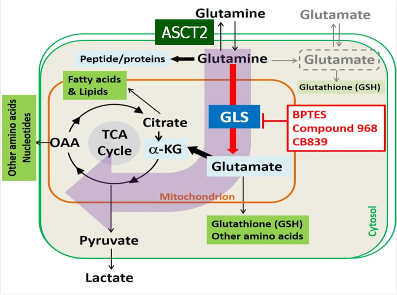
This project aims to develop and validate quantitative analysis tools for [11C]Gln (L-[5-11C]Glutamine) and [18F]Fluoroglutamine ((2S,4R)-4[18F]Glutamine) PET using human breast cancer xenografts.
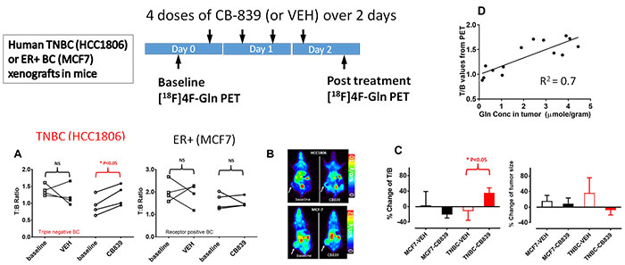
Based on our preliminary studies, we have designed studies to test the central hypothesis that “Distribution volume of [18F]Fluoroglutamine is a marker of tumor glutamine pool that is an indirect measure of glutamine metabolism (Figure 2), while [11C]Gln flux through enzyme glutaminase is an authentic marker of tumor glutaminase activity”.
Figure 2.
Past TMMI projects
Mounting evidence from basic research over the past decade reveals that inflammation and endothelial dysfunction are the central processes in all stages of atherosclerosis; determining levels of the inflammatory mediators (cells such macrophages, or enzymes such as matrix metalloproteinase) accumulated in the plaque would be a way to estimate vulnerability. We have modified human low density lipoprotein (isolated from donor blood) and turned them into nanoparticles carrying a GdDPTA contrast agent. MRI can visualize their uptake by the macrophages in the atheroplaque of apoE-/- mice.
We also developed high-density lipoprotein (HDL)-like nanoparticle that contains iron oxide clusters whose uptake in the atheroplaque is confirmed by Prussian blue staining.
Figure 1.
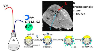
Figure 2.
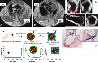
Arteriolosclerosis in LDLr-/- mouse detected by in vivo (A-B) and ex vivo (C) MR imaging. Diagram of lipoprotein-iron oxide (IO) nanoparticles (D) and Prussian blue staining of artery segments after injection of nanoparticles (E). AA= ascending aorta, DA= descending aorta.
Our laboratory is focused on two aspects of stem cell mediated cardiac repair:
- Develop in vivo imaging methods to track the fate (survival, proliferation and differentiation) of stem cells and evaluate their effects on global and regional cardiac function. We have developed magnetic resonance imaging (MRI) methods to measure the global and contractile function in living mice.
- Examine embryonic stem cell (ESC) derived cardiomyocytes (CM) for the treatment of an infarcted heart. Utilizing murine ESCs, carrying an early cardiac specific gene whose expression is linked to an antibiotic resistant gene, we are able to purify the CM population from beating embryoid bodies and obtain sufficient number of cells for in vivo study.
The long-term goal of this project is to facilitate the clinical translation of stem cell mediated therapy for myocardial infarction by identifying the optimal cell type (ESCs, skeletal myoblasts, bone marrow derived or resident cardiac progenitor cells) and understanding the mechanism of their capability to improve the cardiac function.
Figure 1.
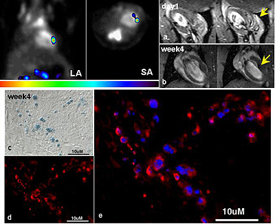
The long (LA) and short (SA) axis view of a rat heart receiving 5 million ESCs, that are stably transfected with a reporter gene (HSV1-sr39tk) and labeled with SPIO particles. [F-18]FHBG signal from grafted cells are in color while myocardial wall is delineated by [N-13] ammonia signal in grey scale. Hypointense region on MR images early after injection represent distribution of cells in the myocardium (a). Over time, the hypointense area reduces while contrast-to-noise ratio remains (b) reflecting the fact that SPIO labels are diluted due to proliferation of ESCs and labels in the dead cells are transferred to macrophages as demonstrated by double staining of SPIO (c) and CD68 (d). The overlay of the two suggests that the majority of SPIO positive cells are infiltrated macrophages.
Figure 2.
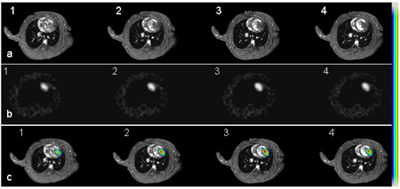
Short axis cardiac MR images of an infarcted heart 14 days after receiving 5M double-labeled ESCs (a, panel 1-4 from base to the apex of the heart). b: [F-18]FHBG images acquired from the same animal. These images have been reformatted to the short axis views as MR images. c: Co-registered PET and MR images using a mutual-information based algorithm. Color scale was used for PET signal while grey scale for MR images.
- Under an approved IRB, explanted hearts are procured from the UPenn Heart Transplantation Program. A myocardial wedge (approx. 2 gram) is dissected from the LV anterior-septal wall and cannulated at ~ 7th branch of the left anterior descending coronary artery (Figure 1). The wedge is perfused with Krebs-Henseleit buffer containing carbon-13 labeled or unlabeled substrates such as glucose, ketone bodies and palmitate. The perfusion media is oxygenated with 95/5 percent O2/CO2 and the pH is maintained at 7.4 ±0.2. The catheterized wedge is secured in a 20 mm NMR tube and then placed inside a 400 MHz NMR spectrometer. High resolution NMR is also performed on tissue extracts.
- Bonded cumulative isotopomers (Bonded Cumomers) mathematical model (Shestov et al. Neurochem Res 11: 2388-401, 2012) is used to derive the quantitative flux measurement by taking advantage of fine-structures carbon-13 MR spectra (singlet, doublet, triplet etc.) and mass balance and isotopomer balance at each (rate-limiting) step of the pathway.
Figure 1.
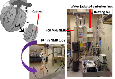
Perfused heart wedges for Carbon-13 NMR-based metabolic studies.
Non-invasive measurement of receptor and gene expression on a low density lipoprotein receptor (LDLr) knockout mouse model, we have developed MR imaging and spec method to evaluate the restoration of LDLR gene expression.
The long term goal of this project is to develop a report-gene approach combined with non-invasive imaging/spectroscopic methods for quantification of transgene expression in the liver, which is a major target organ for gene therapies aimed at correcting metabolic disorder (such as familial hypercholesterolemia) or cancer.
Figure 1.
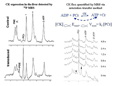
Creatine kinase (CK) expression in the mouse liver detected by in vivo 31P MR spectroscopy (left panel) and quantification of CK concentration (proportional to CK flux) by a progressive saturation transfer experiment (right panel). The phosphocreatine (PCr) resonance only occurs in the liver transfected with creatine kinase (CK), which is not normally expressed in the liver. The ɣ-ATP (arrow head) is saturated for the time indicated on the right side.
Publications
- Young AJ, Pantel AR, Kiani M, Doot RK, Bagheri S, Pryma DA, Farwell MD, Li S, Lee H, Schubert EK, Secreto A, Zuckerman SP, Nayak A, Choi H, Carlin S, DeMichele A, Mankoff DA, Zhou R, Mach RH, McDonald ES. Kinetic Analysis and Metabolism of Poly(Adenosine Diphosphate–Ribose) Polymerase-1–Targeted 18F-Fluorthanatrace PET in Breast Cancer. Journal of Nuclear Medicine 2024;65(12):1862-1868.
- Muller FM, Vervenne B, Maebe J, Blankemeyer E, Sellmyer MA, Zhou R, Karp JS, Vanhove C, Vandenberghe S. Image Denoising of Low-Dose PET Mouse Scans with Deep Learning: Validation Study for Preclinical Imaging Applicability. Mol Imaging Biol 2024;26(1):101-113.
- Li Y, Joaquim MR, Pickup S, Song HK, Zhou R, Fan Y. Learning ADC maps from accelerated radial k-space diffusion-weighted MRI in mice using a deep CNN-transformer model. Magnetic Resonance in Medicine 2024;91(1):105-117.
- Hensley CT, Padakanti P, Damani R, Dulal C, Choi H, Li S, Cao J, Lee H, Pantel A, Li E, Mankoff D, Zhou R. L-5-[11C]-glutamine PET of breast cancer: Preclinical studies in mouse models. Nuclear Medicine and Biology 2025;150-151:109092
- Gupta M, Choi H, Kemp SB, Furth EE, Pickup S, Clendenin C, Orlen M, Rosen M, Liu F, Cao Q, Stanger BZ, Zhou R. Multimetric MRI Captures Early Response and Acquired Resistance of Pancreatic Cancer to KRAS Inhibitor Therapy. Clinical Cancer Research 2025;31(13):2663-2674
- Rong Zhou, Yong Fan, Emma Furth, Ernesto Lima, Thomas Yankeelov: MRI Detection of PDAC Response to KRAS Inhibitor Therapy: Reply-to-Editor. Clinical Cancer Research 31(15): 3345, June 2025.
- Choi H, Gupta M, Sengupta A, Furth EE, Hensley C, Weljie AM, Lee H, Lu Y-T, Pantel A, Mankoff D, Zhou R. Disruption of redox balance in glutaminolytic triple negative breast cancer by inhibition of glutaminase and glutamate export. Neoplasia 2025;61:101136.
- Fracassi A, Qiao H, Lowell AN, Cao J, Bode JW, Masai H, Yoshizawa-Sugata N, Zhou R, Yamakoshi Y. Natural and Synthetic LDL-Based Imaging Probes for the Detection of Atherosclerotic Plaques. ACS Pharmacology & Translational Science 2025; 8: 578-591.
- Pickup S, Romanello M, Gupta M, Song HK, Zhou R. Dynamic Contrast-Enhanced MRI in the Abdomen of Mice with High Temporal and Spatial Resolution Using Stack-of-Stars Sampling and KWIC Reconstruction. Tomography. 2022; 8(5):2113-2128. https://doi.org/10.3390/tomography8050178
- Romanello Joaquim, E.E. Furth, Y. Fan, H.K. Song, S. Pickup, J. Cao, H. Choi, M. Gupta, Q. Cao, R. Shinohara, D. McMenamin, C. Clendenin, T.B. Karasic, J. Duda, J.C. Gee, P.J. O’Dwyer, M.A. Rosen, R. Zhou, DWI Metrics Differentiating Benign Intraductal Papillary Mucinous Neoplasms from Invasive Pancreatic Cancer: A Study in GEM Models, Cancers 2022; 14(16): 4017. doi:10.3390/cancers14164017
- Choi H, Cao J, Qiao H, Chen I-W, Zhou R. Improving Cancer Detection and Treatment by pH-Sensitive Peptide Nanoparticle Drug Delivery Platform: Pharmacokinetics, Toxicity, and Immunogenicity Profile. Advanced NanoBiomed Research 2022;2(3):2100081.
- Zhou R, Choi H, Cao J, Pantel A, Gupta M, Lee HS, Mankoff D. [18]F-Fluciclovine PET Imaging of Glutaminase Inhibition in Breast Cancer Models. J Nucl Med 2023;64(1):131-136.
- Cao J, Pickup S, Rosen M, Zhou R. Impact of Arterial Input Function and Pharmacokinetic Models on DCE-MRI Biomarkers for Detection of Vascular Effect Induced by Stroma-Directed Drug in an Orthotopic Mouse Model of Pancreatic Cancer. Mol Imaging Biol 2023;25(4):638-647
- Malyarenko D, Amouzandeh G, Pickup S, Zhou R, Manning HC, Gammon ST, Shoghi KI, Quirk JD, Sriram R, Larson P, Lewis MT, Pautler RG, Kinahan PE, Muzi M, Chenevert TL. Evaluation of Apparent Diffusion Coefficient Repeatability and Reproducibility for Preclinical MRIs Using Standardized Procedures and a Diffusion-Weighted Imaging Phantom. Tomography : a journal for imaging research 2023;9(1):375-386.
- Moore SM, Quirk JD, Lassiter AW, Laforest R, Ayers GD, Badea CT, Fedorov AY, Kinahan PE, Holbrook M, Larson PEZ, Sriram R, Chenevert TL, Malyarenko D, Kurhanewicz J, Houghton AM, Ross BD, Pickup S, Gee JC, Zhou R, Gammon ST, Manning HC, Roudi R, Daldrup-Link HE, Lewis MT, Rubin DL, Yankeelov TE, Shoghi KI. Co-Clinical Imaging Metadata Information (CIMI) for Cancer Research to Promote Open Science, Standardization, and Reproducibility in Preclinical Imaging. Tomography : a journal for imaging research 2023;9(3):995-1009.
- D.M. Peehl, C.T. Badea, T.L. Chenevert, H.E. Daldrup-Link, L. Ding, L.E. Dobrolecki, A.M. Houghton, P.E. Kinahan, J. Kurhanewicz, M.T. Lewis, S. Li, G.D. Luker, C.X. Ma, H.C. Manning, Y.M. Mowery, P.J. O'Dwyer, R.G. Pautler, M.A. Rosen, R. Roudi, B.D. Ross, K.I. Shoghi, R. Sriram, M. Talpaz, R.L. Wahl, R. Zhou, Animal Models and Their Role in Imaging-Assisted Co-Clinical Trials, Tomography. 2023; 9(1): 657-680.
- Shoghi, C. Badea, S. Blocker, T. Chenevert, R. Laforest, M. Lewis, G. Luker, H. Manning, D. Marcus, Y. Mowery, S. Pickup, A. Richmond, B. Ross, A. Vilgelm, T. Yankeelov, R. Zhou, Co-Clinical Imaging Resource Program (CIRP): Bridging the Translational Divide to Advance Precision Medicine, Tomography 2020: 6(3): 273-287.
- Fracassi A, Cao J, Yoshizawa-Sugata N, Tóth É, Archer C, Gröninger O, Ricciotti E, Tang SY, Handschin S, Bourgeois J-P, Ray A, Liosi K, Oriana S, Stark W, Masai H, Zhou R, Yamakoshi Y. LDL-mimetic lipid nanoparticles prepared by surface KAT ligation for in vivo MRI of atherosclerosis. Chemical Science 2020;11(44):11998-12008
- Du T, Zhang H, Li Y, Pickup S, Rosen M, Zhou R, Song HK, Fan Y. Adaptive convolutional neural networks for accelerating magnetic resonance imaging via k-space data interpolation. Medical Image Analysis. 2021;72:102098.
- Viswanath V, Zhou R, Lee H, Li S, Cragin A, Doot RK, Mankoff DA, Pantel AR. Kinetic Modeling of (18)F-(2S,4R)4-Fluoroglutamine in Mouse Models of Breast Cancer to Estimate Glutamine Pool Size as an Indicator of Tumor Glutamine Metabolism. J Nucl Med 2021;62(8):1154-1162
- Cao J, Song HK, Yang H, Castillo V, Chen J, Clendenin C, Rosen M, Zhou R, Pickup S. Respiratory Motion Mitigation and Repeatability of Two Diffusion-Weighted MRI Methods Applied to a Murine Model of Spontaneous Pancreatic Cancer. Tomography. 2021;7(1):66-79.
Alumni
- Zijun Li
Postdoc Research Training Period: 2003-2004
Prior Academic Degree(s): MD, PhD
Prior Academic Degree Institution: Sun Yatsen University of Medical Sciences - Daniel Thomas
Postdoc Research Training Period: 2003-2005
Prior Academic Degree(s): MD
Prior Academic Degree Institution: University of Cologne - EJ Kim
Postdoc Research Training Period: 2003-2004
Prior Academic Degree(s): PhD
Prior Academic Degree Institution: Yonsei University - Hui Qiao
Research Associate /Laboratory Manager 2004-2010
Prior Academic Degree(s): MD, PhD
Prior Academic Degree Institution: Sun Yat-Sen University of Medical Sciences - Fabao Gao
Postdoc Research Training Period: 2005-2007
Prior Academic Degree(s): MD, PhD
Prior Academic Degree Institution: Fourth Military Medical University - Hoon Choi
Postdoc Research Training Period: 2006-2009
Prior Academic Degree(s): PhD
Prior Academic Degree Institution: Pohang University of Science and Technology - Jia Zhong
Postdoc Research Training Period: 2009-2010
Prior Academic Degree(s): PhD
Prior Academic Degree Institution: Case Western Reserve University - Song Chen
Postdoc Research Training Period: 2011-2012
Prior Academic Degree(s): PhD
Prior Academic Degree Institution: Xiamen University, P.R. China - Luca D'Agostino
Postdoc Research Training Period: 2012-2013
Prior Academic Degree(s): PhD
Prior Academic Degree Institution: University of Siena, Siena, Italy - Ting Liu
Research Training Period: 2012-2014
Prior Academic Degree(s): PhD
Prior Academic Degree Institution:Huaxi Medical University, Sichuan, P.R. China - Mamta Gupta
Research Training Period: 2021-2025
Prior Academic Degree(s): PhD
Prior Academic Degree Institution: Fortis Memorial Research Institute, Gurgaon, Haryana, India.
- Miguel Romanello Joaquim
Training Period (Degree): 2020-2022 (MS, University of Pennsylvania)
Prior Academic Degree(s): BS
Prior Academic Degree Year(s): 2020
Prior Academic Degree Institution: Notre Dame University - Jianbo Cao
Training Period (Degree): 2016- (PhD)
Prior Academic Degree(s): BS, MS
Prior Academic Degree Year(s): 2012 (BS), 2015 (MS)
Prior Academic Degree Institution: Xiamen University, China - Shubutm Islam
Training Period (Degree): 2008 -2012(MS)
Prior Academic Degree(s): BS
Prior Academic Degree Year(s): 2012
Prior Academic Degree Institution: University of Pennsylvania - Robert Jodi
Training Period (Degree): 2010-2011 (Distinction in Research Program)
Prior Academic Degree(s): BS
Prior Academic Degree Year(s): 2008
Prior Academic Degree Institution: Emory University - Ting Liu
Training Period (Degree): Visiting 2011-2012 (PhD)
Prior Academic Degree(s): MD
Prior Academic Degree Year(s): 2011
Prior Academic Degree Institution: Huaxi Medical University -
Shelley (Hualei) Zhang
Training Period (Degree): 2005 -2010(PhD)
Prior Academic Degree(s): BS
Prior Academic Degree Year(s): 2003
Prior Academic Degree Institution: Hong Kong University of Science and Technology
- Ashley B. Bakken
Training Period: 2007
Prior Academic Degree: BA - Anh Nguyen
Training Period: 2013-2015
Academic Institution: University of Pennsylvania - Lovemore S. Kuzomunhu
Training Period: 2013-2014
Academic Institution: University of Pennsylvania
Prior Academic Degree: BS - Harrison Mace
Training Period: 2014
Academic Institution: University of Pennsylvania
Prior Academic Degree: BS - Hana Page
Training Period: 2014-2015
Academic Institution: University of Pennsylvania
Prior Academic Degree: BS - Edward Jin
Training Period: 2015
Academic Institution: University of Pennsylvania - Morgan Venuti
Training Period: 2015
Academic Institution: University of Pennsylvania - Connor G. Hoge
Training Period: 2015-2016
Academic Institution: University of Pennsylvania
Prior Academic Degree: BS - Helen Wang
Training Period: 2016
Academic Institution: Case Western Reserve University - Ritwik Bhatia
Training Period: 2016
Academic Institution: University of Pennsylvania - Daniel Kranseler
Training Period: 2017- 2018
Academic Institution: University of Pennsylvania - Hanwen Yang
Training Period: 2018-2021
Academic Institution: University of Pennsylvania