
INSIGHT Lab
Research
Three fronts, one throughline
Every project in the lab answers the same question at a different stage of translation: can we see what a therapeutic cell is doing, from the manufacturing bench to the patient's tumor?
Molecular Imaging, Nanomedicine & Cell-Based Therapeutics
Team Lead: Maryam Soleimani, Postdoctoral Fellow1. Engineer & label
Mechanoporation loads CAR T cells and macrophages with iron oxide nanoparticles.
2. Cross the barrier
Focused ultrasound briefly opens the blood–brain barrier for delivery.
3. Track with MRI
Susceptibility mapping follows labeled cells noninvasively over time.
4. Guide therapy
Imaging findings feed back into dosing and treatment decisions.
Developing imaging-enabled approaches to understand, engineer, and guide emerging therapies in preclinical models. Our work integrates molecular imaging, nanomedicine, immune-cell engineering, and image-guided therapeutic delivery, with an emphasis on translating biological processes into measurable imaging biomarkers.
- Engineering MRI, PET, and multimodality-visible therapeutic and immune cells
- Nanoparticle-based cell labeling, molecular imaging, and theranostic platforms
- Tracking cellular delivery, biodistribution, persistence, viability, and therapeutic response
- Development of quantitative MRI and susceptibility-based imaging biomarkers
- Image-guided optimization of cellular and molecular therapies
- Preclinical models of brain tumors, neuroinflammation, and other neurologic diseases
- Development of imaging approaches for tumor targeting, margin characterization, and treatment guidance
Translational Imaging & Image-Guided Therapy
Team Lead: Rachel Saks, Former Penn Neuroradiology Fellow and current Neuroradiology Attending at Northwell – Lenox Hill Hospital1. Image the patient
Advanced and quantitative MRI captures disease and therapy in vivo.
2. Quantify biomarkers
Volumetric and susceptibility measures turn images into numbers.
3. Follow response
Serial imaging separates true progression from treatment effect.
4. Guide therapy with new insight
Findings shape treatment planning, targeting, and toxicity monitoring.
Translating emerging imaging technologies from experimental models into clinically meaningful tools for diagnosis, treatment guidance, and therapeutic monitoring. Our clinical research combines advanced neuroimaging, quantitative biomarkers, and longitudinal imaging to better characterize disease biology and evaluate novel therapies.
- Clinical translation of molecular and cellular imaging technologies
- Imaging of immunotherapy, cell therapy, and other emerging therapeutics
- Monitoring therapeutic delivery, treatment response, and treatment-related toxicity
- Quantitative MRI and advanced imaging biomarkers in neuro-oncology
- Longitudinal assessment of brain tumors and treatment effects
- Imaging biomarkers for neurodegenerative, inflammatory, and vascular disorders
- Image-guided approaches for treatment planning, targeting, and therapeutic monitoring
AI-Enabled Imaging Intelligence & Clinical Decision Support
Team Lead: Shahriar Faghani, Radiology Resident1. Gather prior data
Prior studies, reports, and clinical records are pulled together.
2. Register & compare
Serial scans are aligned so interval change stands out.
3. Detect & summarize
Vision and language models flag findings and write longitudinal summaries.
4. Guide diagnosis & therapy
Answers arrive at the radiologist’s workstation, where decisions are made.
Developing AI systems that transform longitudinal imaging and clinical data into actionable information at the radiologist’s workstation. Our work combines large language models, computer vision, quantitative imaging, and multimodal AI to improve interpretation, comparison, workflow, and clinical decision-making.
- AI-generated longitudinal summaries of imaging reports and clinical records
- Automated registration, prior-study comparison, and interval change detection
- Brain metastasis detection and longitudinal assessment across serial MRI
- ARIA-E and ARIA-H detection and surveillance during anti-amyloid therapy
- Differentiation and quantification of post-treatment change and tumor progression
- Integration of radiation therapy dose maps with diagnostic imaging
- Automated extraction and quantification of imaging biomarkers
- Multimodal AI integrating imaging, reports, clinical data, and quantitative imaging
Translation & innovation
Interested in partnering on translational or startup opportunities in imaging-guided cell therapy? Get in touch.
- Patented diagnostic nanoparticle technology for cellular apoptosis imaging.
- Provisional patent covering a novel approach to stem cell differentiation.
- Ongoing interest in translating the lab's mechanoporation cell-labeling and AI imaging-biomarker platforms toward commercial and clinical-stage development.
Lab members
A physician-scientist team spanning postdoctoral, resident, fellow, and undergraduate researchers across all three research fronts.
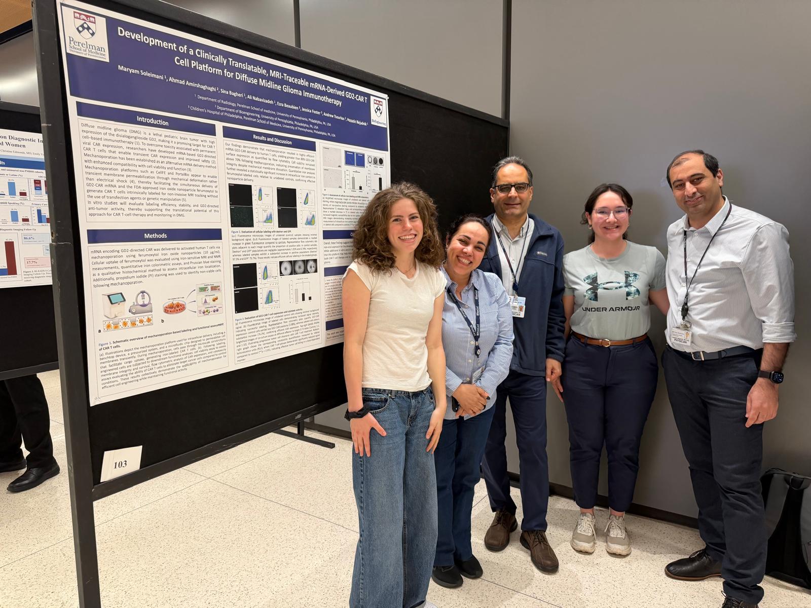
Physician-scientist working at the intersection of molecular imaging, cell engineering, and neuro-oncology. Trained at Stanford's Molecular Imaging Program before joining Penn Radiology.
Preclinical — Immune Cell Therapy & Cell Tracking
Clinical — Immunotherapy Imaging
Informatics - Radiology AI Copilot
Alumni
Lab news
Recent awards, funded grants, and publications from the lab.
McCabe Fund Award supports the lab's imaging-guided cell therapy work
The McCabe Fund Advisory Committee, chaired by Ronald P. DeMatteo, MD, selected the project at its June 29 meeting, citing the spirit of the project and the sound science described. The award runs August 1, 2026 through January 31, 2028.
Early Career Neuroradiology Leadership Development Scholarship, American Society of Neuroradiology
Selected by ASNR for its Early Career Neuroradiology Leadership Development Scholarship.
Outstanding Project Award, American Society of Neuroradiology
Recognized by ASNR for the lab's work on imaging-guided cellular immunotherapy.
Educational Exhibits Award, American Society of Neuroradiology
Honored at the ASNR annual meeting in Austin, TX for an educational exhibit from the lab.
ASNR Foundation funds “Imaging-Guided mRNA GD2-CAR T Cell Therapy” project
A two-year award from the American Society of Neuroradiology Foundation to develop MRI-visible mRNA GD2-CAR T cells for diffuse midline glioma.
RSNA seed grant awarded for neuro-oncology imaging project
RSD26-243 will support noninvasive MRI visualization of therapeutic immune cells in neuro-oncology.
Two abstracts presented at ASNR, Austin, TX
“Nanoparticle-Enhanced MR Imaging of T-Cell Tracking” and “Imaging Neurotoxicity in CAR T-Cell Therapy: A Neuroradiologist's Perspective.”
Abstract presented at the European Congress of Radiology, Vienna
“Non-Invasive Iron Oxide Labeled T-Cell Tracking via MR Imaging.”
In Vivo Imaging of Nanoparticle-Labeled CAR T Cells published in PNAS
Kiru et al. demonstrate the first in vivo imaging of nanoparticle-labeled CAR T cells, a foundation for the lab's current CAR-T imaging work.
Mechanoporation radiolabeling method published in Scientific Reports
Jung et al. show mechanoporation enables rapid, efficient PET radiolabeling of stem cells.
Selected publications
Full publications list available on Google Scholar.
Join the lab or collaborate with us.
We welcome inquiries from postdoctoral fellows, residents, fellows, and undergraduate researchers, as well as collaborators in imaging, immunotherapy, and AI — and investors or partners interested in our translational work.
Three positions are currently open; explore the open positions below for full descriptions.
- Email: Hossein.Nejadnik@PennMedicine.UPenn.edu
- Department: Department of Radiology, Hospital of the University of Pennsylvania, 3400 Spruce Street, Philadelphia, PA 19104
- Lab: Department of Bioengineering, 435S Skirkanich Hall, 210 S. 33rd Street, Philadelphia, PA 19104
- ORCID: orcid.org/0000-0003-1249-0724
Open positions
We are actively recruiting across all three research fronts. Applications are reviewed on a rolling basis; start dates are flexible.
Postdoctoral Fellow — CAR T-Cell Therapy, Cancer Biology, and Molecular Imaging
Full-time postdoctoral position · University of Pennsylvania, Philadelphia, PA
The Nejadnik Laboratory is seeking a highly motivated postdoctoral fellow to join an interdisciplinary program developing next-generation CAR T-cell therapies for pediatric brain tumors. The work integrates cellular immunotherapy, molecular imaging, nanotechnology, and translational cancer biology, currently focused on GD2-directed CAR T cells for diffuse midline glioma, including H3K27M-mutant tumors, with an emphasis on rapid CAR T-cell manufacturing, noninvasive tracking of therapeutic cells, and imaging-guided optimization of treatment and repeat-dose strategies.
Full position description
Project areas:
- Engineering and characterization of CAR T cells, including transient mRNA-based CAR expression
- Primary human T-cell culture, activation, transfection, and functional assessment
- Flow cytometry analysis of CAR expression, viability, activation, memory, and exhaustion phenotypes
- In vitro tumor-cell killing and cytokine-release assays
- Development and use of orthotopic mouse models of diffuse midline glioma
- Intravenous and intracranial delivery of therapeutic cells
- Longitudinal assessment of CAR T-cell trafficking, persistence, antitumor response, and safety
- Molecular and cellular imaging, including bioluminescence imaging and MRI-based cell tracking
- Histology, immunohistochemistry, and imaging–pathology correlation
- Preparation of manuscripts, scientific presentations, and grant-related materials
Required qualifications:
- PhD, MD, MD/PhD, or equivalent doctoral degree in immunology, cancer biology, cell and molecular biology, biomedical engineering, neuroscience, bioengineering, or a related field
- Strong background in experimental biomedical research
- Experience with mammalian cell culture and standard molecular or cellular biology techniques
- Ability to work independently, analyze data rigorously, and collaborate within a multidisciplinary team
- Strong scientific writing and communication skills
Preferred qualifications:
- CAR T-cell engineering or adoptive cellular immunotherapy
- Primary human T-cell isolation, culture, activation, and functional characterization
- Flow cytometry and multicolor immunophenotyping
- Preclinical cancer models and small-animal experimentation
- Orthotopic brain-tumor models, stereotactic surgery, or intracranial cell delivery
- Bioluminescence imaging, IVIS, MRI, quantitative imaging, or image analysis
- Iron oxide nanoparticles, cell labeling, or molecular imaging
- Histology, immunofluorescence, or immunohistochemistry
- mRNA delivery, transfection, electroporation, microfluidics, or mechanoporation
- Quantitative data analysis and statistical programming
Candidates are not expected to have experience in every area. Applicants with a strong background in CAR T-cell biology and an interest in developing animal-model and imaging expertise are encouraged to apply, as are candidates with strong preclinical animal and imaging experience who wish to expand into cellular immunotherapy.
Research environment: The fellow will work in a highly collaborative environment spanning radiology, bioengineering, immunology, oncology, neuroscience, and small-animal imaging, with mentorship in experimental design, scientific writing, manuscript and grant preparation, presentations, and career advancement.
How to apply: Cover letter describing research background, technical experience, and interest in the position; curriculum vitae; contact information for three professional references; and one or two representative publications.
Email materials to Hossein.Nejadnik@PennMedicine.UPenn.edu with the subject line “Postdoctoral Application – CAR T-Cell Therapy.”
Postdoctoral Research Fellow — Artificial Intelligence in Medical Imaging and Healthcare
Department of Radiology · Perelman School of Medicine · Philadelphia, PA (hybrid arrangements may be considered)
We are seeking a postdoctoral research fellow to help build the Radiology AI Copilot — a program at the intersection of large language models, multimodal AI, medical image analysis, and clinical decision support. Two threads run through the work: AI summarization of imaging reports and the clinical record, and an image-processing effort centered on automated comparison of current studies against priors.
Full position description
Research areas:
- AI summarization — LLM- and RAG-based summarization of longitudinal radiology reports and clinical data
- Current-vs-prior image comparison — registration, subtraction, and change detection across serial studies, applied to brain tumor metastasis detection, ARIA-E/ARIA-H monitoring in anti-amyloid therapy, FLAIR signal abnormality (post-treatment change vs. progression), and radiation dose-map overlay onto diagnostic MRI
- Multimodal AI combining imaging, text, and clinical data
- Medical image analysis using deep learning and foundation models
- Clinical decision support systems and radiology workflow optimization
- Human–AI interaction and evaluation of AI systems in clinical environments
- Imaging informatics and health services research
The fellow will have substantial opportunity to develop independent research directions aligned with their interests and career goals.
Responsibilities:
- Develop, train, and evaluate machine learning and AI models for healthcare applications
- Design and implement LLM-based and multimodal AI systems
- Analyze large-scale imaging and clinical datasets
- Conduct experimental studies and model evaluations
- Prepare manuscripts, conference abstracts, and grant applications
- Collaborate with clinicians, scientists, engineers, and trainees across disciplines
Required qualifications:
- PhD, MD, MD/PhD, or equivalent doctoral degree in computer science, biomedical informatics, electrical engineering, bioengineering, data science, computational biology, medical physics, or a related field
- Strong programming skills in Python
- Experience with deep learning frameworks such as PyTorch or TensorFlow
- Demonstrated research productivity through peer-reviewed publications
- Excellent communication and teamwork skills
Preferred qualifications:
- Large language models and generative AI; retrieval-augmented generation (RAG) systems
- Multimodal foundation models and vision-language models
- Medical image analysis, computer vision, and image registration
- Clinical NLP
- Reinforcement learning or agentic AI systems
- Evaluation and deployment of AI systems in healthcare settings
Research environment, compensation, and benefits: Access to extensive clinical imaging datasets and high-performance computing resources; collaborations across radiology, oncology, neuroscience, informatics, and engineering; competitive salary commensurate with experience and full University benefits; professional development and conference travel support.
Application materials: Curriculum vitae; cover letter describing research interests and career goals; contact information for three references; up to three representative publications.
Email materials to Hossein.Nejadnik@PennMedicine.UPenn.edu with the subject line “Postdoctoral Application – AI in Medical Imaging.” Start date flexible; applications reviewed on a rolling basis.
Clinical Research Opportunities in Cellular Therapy, Molecular Imaging, and Neuro-Oncology
Flexible duration · University of Pennsylvania, Philadelphia, PA · Fellows, Residents & Medical Students
We welcome motivated fellows, residents, and medical students interested in clinical and translational research at the intersection of cellular therapy, advanced imaging, and oncology — including pediatric and adult brain tumors, image-guided cellular therapy, MRI-based treatment assessment, and translation of laboratory discoveries into clinically relevant applications.
Full position description
Potential research activities:
- Clinical and imaging database development
- Retrospective and prospective clinical research studies
- Review of electronic medical records and extraction of clinical outcomes
- MRI evaluation of tumor response, treatment-related changes, and neurotoxicity
- Quantitative imaging and imaging-biomarker analysis
- Correlation of imaging findings with treatment, pathology, laboratory, and survival data
- Research involving CAR T-cell therapy and other cellular immunotherapies
- Literature reviews, systematic reviews, and meta-analyses
- Abstract preparation for national meetings; manuscript preparation and publication
- Development of research protocols and IRB submissions
- Collaboration with investigators in radiology, oncology, neuro-oncology, immunology, pathology, and bioengineering
Who should apply: Clinical fellows interested in cellular therapy, oncology, neuro-oncology, radiology, or molecular imaging; residents seeking a focused clinical or translational research project; medical students interested in research experience, scientific meetings, and peer-reviewed publications; and trainees considering careers in academic medicine, radiology, oncology, biomedical imaging, or translational research.
Previous research experience is helpful but not required. Applicants should be dependable, intellectually curious, attentive to detail, and able to commit consistent time to completing an assigned project.
Mentorship and scholarly opportunities: Each trainee is matched with a defined project appropriate for their background and available time, with mentorship in formulating a research question, study design and methodology, clinical and imaging data analysis, interpretation of findings, abstract and manuscript preparation, scientific presentation, and career development in academic medicine.
Time commitment: Projects range from focused short-term studies to longitudinal research experiences. Applicants should indicate whether they are seeking a medical-student research experience, a resident research or scholarly project, a fellowship research project, a research-year opportunity, or a longitudinal mentorship experience.
How to apply: Brief statement describing clinical background, research interests, and career goals; current curriculum vitae or résumé; training level and institutional affiliation; expected availability and preferred duration of involvement; and any relevant experience with clinical research, imaging, statistics, or scientific writing.
Email materials to Hossein.Nejadnik@PennMedicine.UPenn.edu with the subject line “Clinical Research Opportunity – [Fellow/Resident/Medical Student].”
Opportunities are subject to project availability, institutional requirements, appropriate research approvals, and the trainee's ability to make a meaningful time commitment. The University of Pennsylvania is an equal opportunity employer; final postings are subject to applicable University policies and official hiring requirements.