
Abdominal Imaging Fellowship
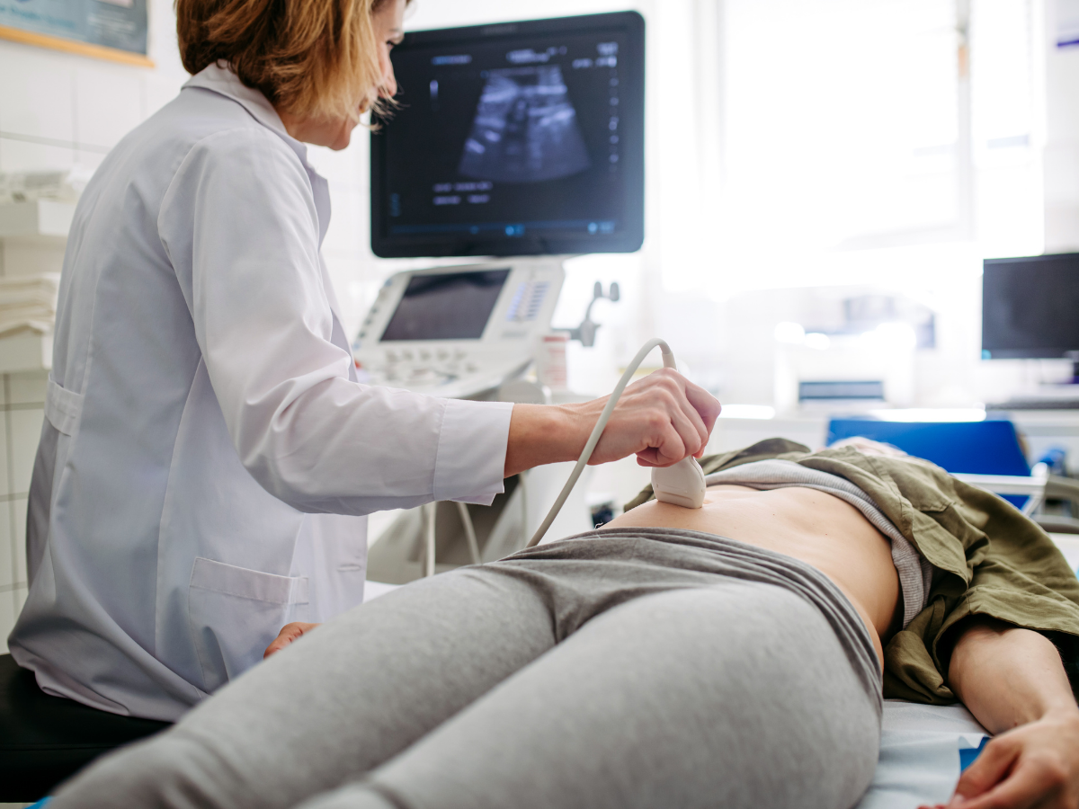
Accredited by the Accreditation Council for Graduate Medical Education (ACGME), our fellows have established highly successful careers in academic and private practices. Up to nine fellows train with us each year. The large size of our program allows for each individual fellow to personalize their educational experience through the use of elective time and by close mentorship from our outstanding faculty. We invite you to continue your radiology training with Penn Radiology, one of the nation's top-ranked radiology departments.
Our goal is to train productive, confident body imagers, who will attain the highest skills in diagnostic interpretation and reporting, enabling them to serve as valued specialty consultants.
Interested in applying?
Learn about program requirements and how to apply, and continue to the sections below to learn more about the program.
Leadership
Matthew A Morgan, MD
Program Director
Contact us
Coordinator: Rachel Alisio
Department of Radiology
Division of Abdominal Imaging
Hospital of the University of Pennsylvania
3400 Spruce Street/1 Silverstein
Philadelphia, PA 19104
Contact: Rachel.Alisio@pennmedicine.upenn.edu | 215-662-3046
Learn more
The Abdominal Imaging Radiology Fellowship program’s principal objectives aim to:
- Expose each fellow to a wide range of pathology across all imaging modalities
- Emphasize the importance of protocol design in optimizing imaging exams
- Discuss the latest evidence-based guidelines for the management of imaging findings
- Teach the technical skills required to perform image-guided biopsies
- Teach trainees a system of self-learning that will allow them to continuously adapt to an ever-changing field of medical science
This one-year course of training consists of 13 blocks, each four weeks long. A typical fellowship year consists of the following rotations:
- 5 blocks on MRI (including subspecialty tumor board assignments with attending)
- 3 blocks on Ultrasound
- 3 blocks on Body CT
- 1 block elective time
- 0.5 blocks of image-guided biopsy and colonography
Each section combines a broad array of didactic teaching with high-volume clinical learning opportunities. The fellows' educational experience also includes approximately five to seven small group seminars per month, presented by the faculty in their subspecialty area of expertise and divisional monthly journal club. The fellows also are active participants in a wide range of multidisciplinary conferences. There is also academic time for fellow research.
Clinical and Research Interests
- Optimization of helical and multidetector scanning techniques
- Helical and multidetector noise and artifacts
- Major clinical projects, including work in appendicitis, inflammatory bowel disease, renal mass characterization, and outcomes research.
Fellowship CT Rotation Goals
- Develop proficiency in the interpretation of CT studies of the abdomen and pelvis, incorporating the following subspecialty areas: gastrointestinal CT, genitourinary CT, oncologic CT, and acute abdominal conditions/trauma.
- Learn the indications and technique of CT-guided percutaneous needle biopsy
- Analyze and evaluate clinical research in body CT
- Learn new techniques in body CT, including 3D analysis of cases using an independent 3D workstation, CT Urography, CT Colonography, CT Enterography, and Dual Energy Body CT
- Develop the ability to function as a teacher and mentor to radiology residents in training
Equipment/Exams
- Over 13,000 clinical MR exams/year at HUP, Penn Presbyterian Medical Center and our community radiology centers
- 8 clinical 1.5T scanners including Siemens Avanto, Essenza, Espree and General Electric 1.5 T optima systems
- 4 clinical whole body 3.0T scanners including two Siemens Verio, Skyra and Tim Trio systems
- 3 whole-body research magnets 1.5, 3.0 and 7T Siemens Avanto, Tim Trio and Magnetom, respectively
- Multi-monitor Sectra PACS workstations
Clinical and Research Interests
- Clinical applications, coil development and technique optimization for abdominal and pelvic MRI
- Clinical research interests include prostate MR, female pelvis, liver, adrenal and renal MR, and use of MR elastography vs US elastography
Fellowship MR Rotation Goals
- Understand the most recent technologic advances and the diagnostic capabilities of MRI
- Establish criteria for patient selection and the appropriate use of MRI
- Incorporate both established and newer MRI techniques into their practice
- Improve their utilization of MRI for numerous clinical indirections
- Learn the latest advances in specialized MRI techniques such as diffusion-weighted images, MR elastography and dynamic contrast enhanced imaging
On the ultrasound (US) rotation, abdominal imaging fellows learn to:
- Interpret high-resolution US findings to evaluate many different types of abdominal pathology
- Systematically use and interpret color Doppler to answer questions in abdominal imaging cases
- Apply new techniques, such as contrast-enhanced and 3D ultrasound
- Understand how to avoid mistakes for emergent conditions, such as ectopic pregnancy, pelvic inflammatory disease, scrotal torsion and acute cholecystitis
Fellows will also learn protocols for performing ultrasound-guided interventional procedures, such as liver mass biopsy, thyroid nodule biopsy, and percutaneous nephrostomy.
Vascular examinations with color, power and duplex Doppler sonography are frequently performed. These are used to assess liver transplants, TIPS evaluation, portal hypertension, renal transplants, impotence, extremities, scrotal and adnexal masses.
We perform more than 18,000 ultrasound examinations annually.