
Penn Radiology Division of Cardiovascular Imaging
Clinical
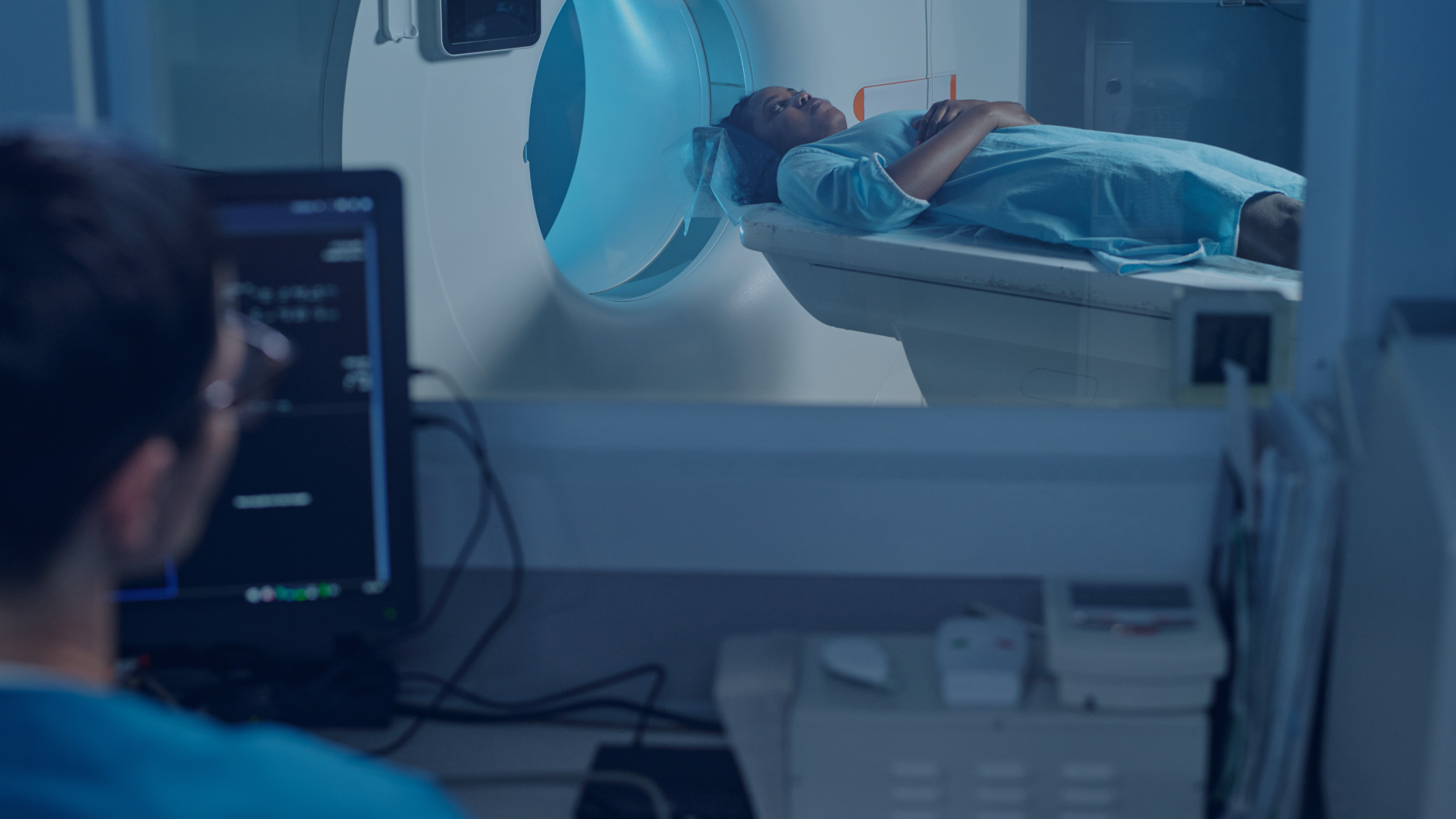
The division continues to practice at the cutting edge by using the latest technology, such as the first dual x-ray source CT scanner, the latest generation of 1.5 and 3TMR systems, and advanced image processing systems, including a virtual reality system for medical image evaluation.
Education
The CVI educational program includes a medical student elective, a mentoring program for biomedical engineering undergraduate and graduate student research, radiology resident and cardiovascular medicine fellow training, and a dedicated one year CVI fellowship, currently with two fellows per year.
Many Penn Radiology residents have chosen to work on research projects in the CVI division, leading to numerous presentations and awards at national and international meetings, grants, and publications.
The Cardiac MR and CT Training program includes semi-annual week-long didactic courses and an ongoing three-month , designed to allow those already in practice to obtain the qualifications necessary to perform and interpret cardiovascular CT and MR.
Research
The division has an active research program, ranging from basic MR pulse sequence development and mathematical modeling of cardiac function, to animal models of atherosclerosis and myocardial infarction, phase I/II trials of novel therapeutics for heart failure and myocardial infarction, novel image reconstruction and post-processing techniques, and clinical studies of the efficacy of new CV imaging techniques.